Ct Anatomy Pelvis Muscles - Abdominal CT anatomy | Radiology Key - The lateral superficial muscles, the transversus and external and internal oblique muscles, originate on the rib cage and on the pelvis (iliac crest and inguinal ligament) and are attached to the anterior and posterior layers of the sheath of the rectus.
byAdmin•
0
Ct Anatomy Pelvis Muscles - Abdominal CT anatomy | Radiology Key - The lateral superficial muscles, the transversus and external and internal oblique muscles, originate on the rib cage and on the pelvis (iliac crest and inguinal ligament) and are attached to the anterior and posterior layers of the sheath of the rectus.. Anatomical drawing of the female pelvis. The lateral superficial muscles, the transversus and external and internal oblique muscles, originate on the rib cage and on the pelvis (iliac crest and inguinal ligament) and are attached to the anterior and posterior layers of the sheath of the rectus. 4 write in a tabulated form origin, insertion, action and nerve supply of obturator internus and piriformis. Muscles of the pelvis that cross the lumbosacral joint to attach onto the trunk were described in the previous blog post note: If you are a pelvic health professional who is looking to reach more.
See more ideas about anatomy, pelvic floor, pelvic floor dysfunction. Hint you are sitting on it right now. 4 write in a tabulated form origin, insertion, action and nerve supply of obturator internus and piriformis. The lateral superficial muscles, the transversus and external and internal oblique muscles, originate on the rib cage and on the pelvis (iliac crest and inguinal ligament) and are attached to the anterior and posterior layers of the sheath of the rectus. As such you can also divide the musculature that moves the thigh at the hip.
Presentation1.pptx, ct normal anatomy of the abdomen and ... from image.slidesharecdn.com Renal pelvis or ureter cancer. Axial pelvis ct axial femur ct axial femur ct axial knee ct. Differences between the male pelvis and the female pelvis. If you want to learn how to read ct scans of the abdomen and pelvis proficiently, this video is an excellent starting point. We created an anatomical atlas of abdominal and pelvic ct which is an interactive tool for studying the conventional anatomy of the normal structures based on a multidetector computed tomography. Pelvic floor muscles that are located wholly within the pelvis. Male abdomen and pelvis ct scan form no 7. Axial mr high resolution (small fov).
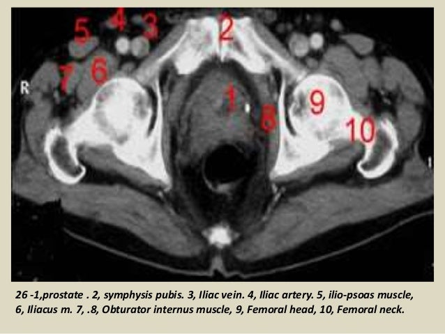
Axial section through male bladder.
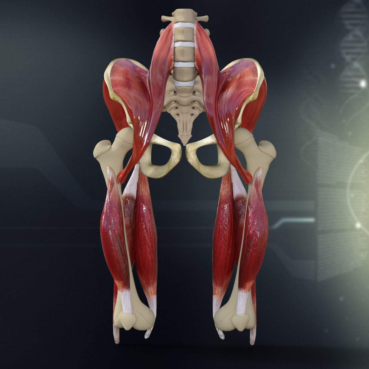
Hepatocellular carcinoma or liver cancer. These muscles origin in continuity from the body of the pubis, along a tendinous arch over the obturator internus fascia, and the ischial spine. Muscles of the pelvis that cross the lumbosacral joint to attach onto the trunk were described in the previous blog post note: The bony pelvis muscles and ligaments figs 6167 the pelvis fig. They support the pelvic organs especially during increases in intra abdominal pressure and also aid in urinary and faecal. This article reviews the anatomical and functional information of the gastrocnemius muscle, its embryological derivation. Anatomy of the thorax, heart, abdomen and pelvis the following video will go through normal abdominal anatomy on ct imaging. The lateral superficial muscles, the transversus and external and internal oblique muscles, originate on the rib cage and on the pelvis (iliac crest and inguinal ligament) and are attached to the anterior and posterior layers of the sheath of the rectus. These muscles, including the gluteus maximus and the hamstrings, extend the thigh at the hip in support of the body's weight and propulsion. Attached to the pelvis are muscles of the buttocks, the lower back, and the thighs. Anatomy pelvis muscles pubococcygeus, puborectalis and iliococcygeus., pelvis nerve, the spinal nerves that arise from vertebral column through the sacrum., pelvic floor musculature laminated anatomy anatomy pelvis muscles; Use the mouse scroll wheel to move the images up and down alternatively use the tiny arrows (>>) on both side of the image to move the images. The muscles of the pelvis form its floor.
Muscles of the pelvis that cross the lumbosacral joint to attach onto the trunk were described in the previous blog post note: The gastrocnemius muscle is a complex muscle that is fundamental for walking and posture. Pelvic floor muscles that are located wholly within the pelvis. As such you can also divide the musculature that moves the thigh at the hip. Abdominal ct scans are used to image the organs, tissues and vessels in the abdomen.
Human Pelvis Muscle Bone Anatomy 3D model | CGTrader from img2.cgtrader.com We created an anatomical atlas of abdominal and pelvic ct which is an interactive tool for studying the conventional anatomy of the normal structures based on a multidetector computed tomography. Renal pelvis or ureter cancer. These muscles origin in continuity from the body of the pubis, along a tendinous arch over the obturator internus fascia, and the ischial spine. Labeled scrollable mri of the pelvis covering anatomy with a level of detail appropriate for medical students. This article reviews the anatomical and functional information of the gastrocnemius muscle, its embryological derivation. 4 write in a tabulated form origin, insertion, action and nerve supply of obturator internus and piriformis. Innervation of the female levator ani muscles. Anatomy of the thorax, heart, abdomen and pelvis the following video will go through normal abdominal anatomy on ct imaging.
Anatomical drawing of the female pelvis.
The full bladder displaces small bowel loops superiorly. They support the pelvic organs especially during increases in intra abdominal pressure and also aid in urinary and faecal. Ct anatomy of the pelvis. As such you can also divide the musculature that moves the thigh at the hip. If you want to learn how to read ct scans of the abdomen and pelvis proficiently, this video is an excellent starting point. Anatomical structures of the abdomen and pelvis are visible as interactive labeled images. It affects the entire lower limb and the movement of the hip and the lumbar area. Renal pelvis or ureter cancer. 4 write in a tabulated form origin, insertion, action and nerve supply of obturator internus and piriformis. Anatomical drawing of the female pelvis. Learn about anatomy muscles pelvis with free interactive flashcards. Muscles of the pelvis that cross the lumbosacral joint to attach onto the trunk were described in the previous blog post note: We created an anatomical atlas of abdominal and pelvic ct which is an interactive tool for studying the conventional anatomy of the normal structures based on a multidetector computed tomography.
If you are a pelvic health professional who is looking to reach more. Axial section through male bladder. Muscles of the pelvis that cross the lumbosacral joint to attach onto the trunk were described in the previous blog post note: This mri male pelvis axial cross sectional anatomy tool is absolutely free to use. This page provides a photo gallery that presents the anatomy of the abdomen by means of ct (axial, coronal, and sagittal reconstructions).
Hip joint from image.slidesharecdn.com The bony pelvis muscles and ligaments figs 6167 the pelvis fig. This article reviews the anatomical and functional information of the gastrocnemius muscle, its embryological derivation. The muscles of the pelvis form its floor. Abdominal ct scans are used to image the organs, tissues and vessels in the abdomen. The floor of the pelvis is formed by the two muscles named levator ani and coccygeus. The gastrocnemius muscle is a complex muscle that is fundamental for walking and posture. Innervation of the female levator ani muscles. Anatomical drawing of the female pelvis.
Ct anatomy of the pelvis.
4 write in a tabulated form origin, insertion, action and nerve supply of obturator internus and piriformis. Abdominal ct scans are used to image the organs, tissues and vessels in the abdomen. Anatomical structures of the abdomen and pelvis are visible as interactive labeled images. This mri male pelvis axial cross sectional anatomy tool is absolutely free to use. The bony pelvis muscles and ligaments figs 6167 the pelvis fig. It affects the entire lower limb and the movement of the hip and the lumbar area. Attached to the pelvis are muscles of the buttocks, the lower back, and the thighs. We'll go through the on this image, we can also see some of the muscles that we talked about specifically the slowest. The gastrocnemius muscle is a complex muscle that is fundamental for walking and posture. 3 enumerate the muscles of true pelvis. Hint you are sitting on it right now. The fracture line shows how a posterior column fracture runs on the right you see a sagittally reformatted ct (oriented the same way as the anatomic drawing to. If you are a pelvic health professional who is looking to reach more.
Architectural differences in the bony pelvis of women with and without pelvic floor disorders anatomy muscles pelvis. Anatomical structures of the abdomen and pelvis are visible as interactive labeled images.